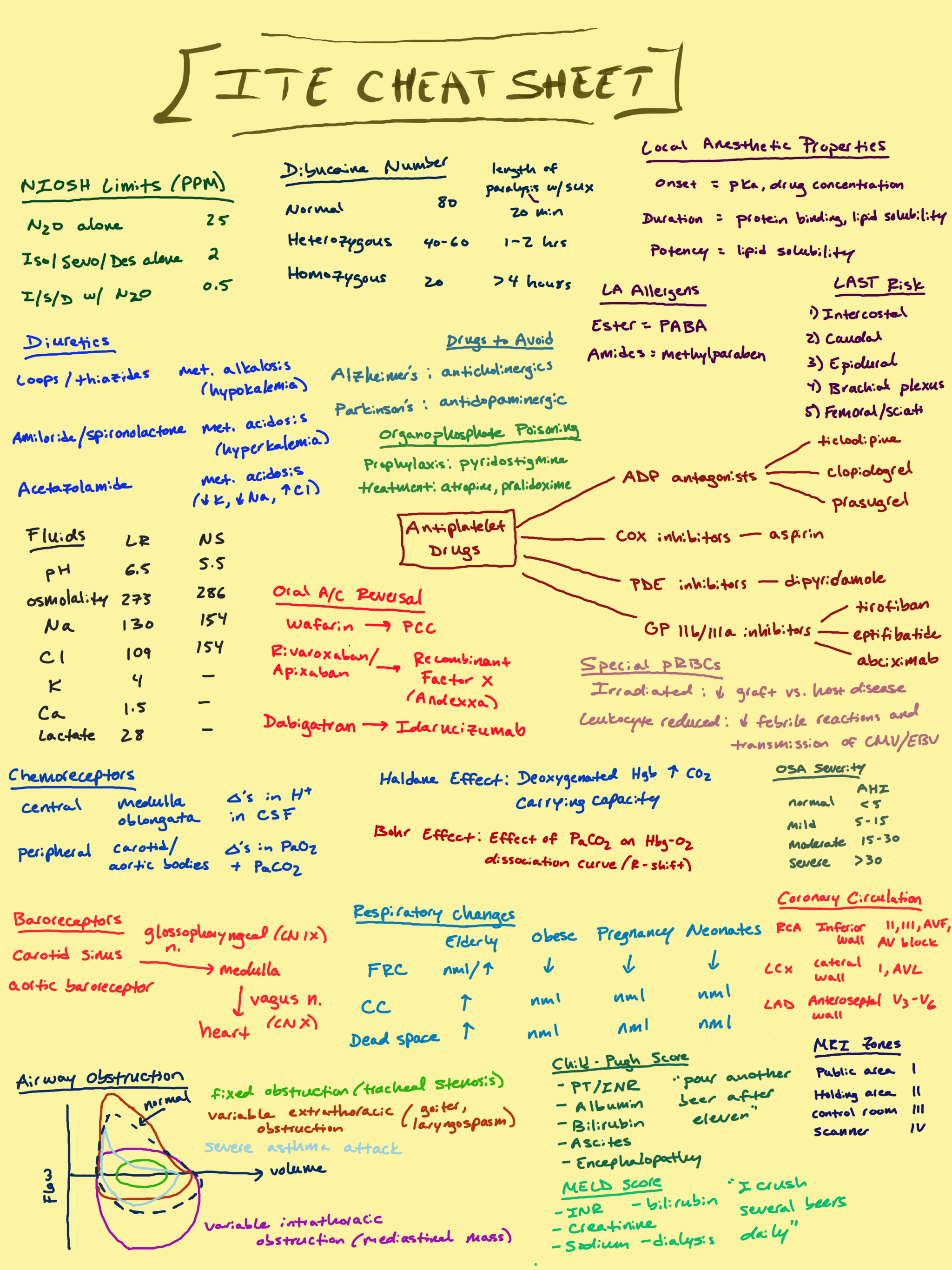
ITE Cram Sheets
The ITE (or In-Training Exam) is an annual exam for Anesthesiology trainees that tests their clinical knowledge. Residency programs typically use it as a benchmark for likelihood of passing the BASIC and ADVANCED Board Exams. These two sheets are designed to be high-yield cram sheets of factoids that I felt like were commonly tested and easy to memorize for the test.
EQUATIAONS
Vaporizer Output = Common Gas Flow (CGF) x SVP/(BP - SVP)
SVP = saturated vapor pressure
BP = barometric pressure
Sevo SVP = 160 mmHg
Iso SVP = 240 mmHg
Des SVP = 669 mmHg
% delivered (dial @ 1 %) = SVP wrong agent/SVP wrong vaporizer
% delivered @ high altitude = 760 mmHg/BP @ high altitude
Airway Resistance = P(peak) - P(plat)/Flow rate
P(peak) = peak pressure
P(plat) = plateau pressure
Maximum Allowable Blood Loss (MABL) = EBV x (start HCT - target HCT)/start HCT
EBV = estimated blood volume
Hct = Hematocrit
Standard Error of Mean = standard deviation/√n
n = number of participants in study
Sensitivity = TP/(TP + FN)
TP = true positive
FN = false negative
Screen Tests
Specificity = TN/(TN+FP)
TN = true negative
FP = false positive
Diagnostic Tests
PPV = TP/(TP+FP)
PPV = positive predictive value
NPV = TN/(TN+FN)
NPV = negative predictive value
Volatiles
Sevoflurane
SVP = 160 mmHg
Blood:Gas coefficient = 0.65
MAC = 2.2
Isoflurane
SVP = 240 mmHg
Blood:Gas coefficient = 1.46
MAC = 1.1
Desflurane
SVP = 669 mmHg
Blood:Gas 0.42
MAC 6.6
Estimated Blood Volumes
Premature 100 ml/kg
Full term 90 ml/kg
Infant 80 ml/kg
Child 75 ml/kg
Male Adult 70 ml/kg
Female Adult 65 ml/kg
Types of Error
Type 1 Error = False Positive
P value 0.05 = 5% chance of type 1 error
Type 2 Error = False negative
Flow Meters
Lower Part —> Laminar Flow —> Affected by viscosity
Upper Part —> Turbulent Flow —> Affected by density
Ultrasound
High Frequency Waves
short wavelength
higher image resolution
limited depth of penetration
Low Frequency Waves
large wavelength
low image resolution
deeper depth of penetration
Color Doppler (“BART”)
Blue = Away from transducer
Red = Towards the transduce
Sympathetic Blocks for Cancer Pain
Stellate Ganglion Block
Head, neck, upper extremity block
Side effects: Horner’s syndrome, increase temperature in upper extremities
Celiac Plexus Block
Upper abdomen, pancreatic block
Side effects: diarrhea, orthostatic hypotension
Superior Hypogastric Block
Pelvic organ block
Lumbar Plexus Block
Low limb block
Airway Nerves
Nasopharynx, Soft/Hard Palate
CN V (maxillary branch)
Anterior 2/3 of Tongue
CN V (mandibular branch)
Posterior 1/3 of Tongue, Pharynx, Superior Epiglottis
Glossopharyngeal nerve
Blocked by tonsillar fossa injection
Inferior Epiglottis, Larynx up to Vocal Cords
Superior Laryngeal nerve
Blocked by greater cornu of hyoid bone injection
Larynx Inferior to Vocal Cords
Recurrent Laryngeal nerve
Blocked by transtracheal injectino
Orthopedic Injuries Leading to Damaged Nerves
Proximal Humerus Fracture
Axillary nerve injury
Mid-Shaft Humerus Fracture
Radial nerve injury
Posterior to Medial Epicondyle (“funny bone”)
Ulnar nerve injury
Anterior Cubital Fossa
Median nerve injury
Spinal Cord Anatomy
Adult Anatomy
Cord ends at L1
Dural Sac ends at S1-2
Neonatal Anatomy
Cord ends at L3
Dural Sac ends at S3-4
NIOSH (National Institute of Occupational Safety and Health) Limits
N20 alone = 25 ppm
Iso/Sevo/Des alone = 2 ppm
Iso/Sevo/Des + N2O = 0.5 ppm
Dibucaine Number (Pseudocholinesterase Deficiency Test)
Normal = 80
length of paralysis with succinylcholine = 20 min
Heterozygous Trait = 40-60
length of paralysis = 1-2 hours
Homozygous Trait =20
length of paralysis = > 4 hours
Local Anesthetics
Properties
onset = pKA, drug concentration
duration = protein binding
potency = lipid solubility
LA Allergens
Ester = PABA
Amides = methylparaben
LAST Risk
Intercostal
Caudal
Epidural
Brachial Plexus
Femoral Sciatic
Drugs to Avoid
Alzheimer’s Disease - avoid anticholinergic drugs
Parkinson’s Disease - avoid antidopaminergic drugs
Organophosphate Poisoning
Prophylaxis = pyridostigmine
Treatment = atropine, pralidoxmine
Diuretic Metabolic Derangement
Loop Diuretics/Thiazides
metabolic alkalosis (hypokalemia)
Amiloride/Spironolactone
metabolic acidosis (hyperkalemia)
Acetazolamide
metabolic acidosis
decreased K+, decreased Na+, increased Cl-
IV Fluids
Lactated Ringers
pH = 6.5
osmolality = 273
Na = 130
Cl = 109
K = 4
Ca = 1.5
Lactate = 28
Normal Saline (NaCl 0.9%)
pH = 5.5
osmolality = 286
Na = 154
Cl = 154
Antiplatelet Drugs
ADP Antagonists
Ticlodipine
Clopidogrel
Prasugrel
COX Inhibitors
Aspirin
PDE Inhibitors
Dipyridamole
GP IIb/IIIa Inhibitors
Tirofiban
Eptifibatide
Abciximab
Oral Anticoagulation Reversal
Warfarin —> PCC
Rivaroxaban/Apixaban —> Recombinant Factor X (Andexxa)
Dabigatran —> Idarucizumab
Specialized Packed RBCs
Irradiated pRBCs
Good for immunodeficient patients
Reduces Grafts vs Host reaction
Leukocyte Reduced
Decreases febrile reactions and transmission of CMV/EBV
Obstructive Sleep Apnea Severity
Acute Hypoxic Index (AHI) Scores
Normal = < 5
Mild = 5-15
Moderate 15-30
Severe > 30
Carbon Dioxide Physics Principles
Haldane Effect
deoxygenated hemoglobin has increased CO2 carrying capacity (venous blood)
Bohr Effects
effect of PaCO2 on hemoglobin-oxygen dissociation curve (rightward shift)
Ventilation Chemoreceptors
Central
medulla oblongata
responds to changes in H+ in CSF
Peripheral
carotid/aortic bodies
responds to changed in PaO2 and PaCO2
Baroreceptors
Carotid sinus/aortic baroceptor —> glossopharyngeal nerve (CN IX) —> medulla —> vagus nerve (CN X) —> heart rate change
Lung Capacity Changes
Elderly
FRC = normal or increased
Closing Capacity = increased
Dead space = increased
Obese
FRC = decreased
CC = normal
Dead space = normal
Pregnancy
FRC = decreased
CC = normal
Dead space = normal
Neonates
FRC = decreased
CC = normal
Dead space = normal
Coronary Circulation
Right Coronary Artery (RCA)
supplies inferior wall
EKG changes on II, III, aVF
can lead to AV block
Left Circumflex Artery (LCX)
supplies lateral wall
EKG changes on I, aVL
Left Anterior Descending (LAD)
supplies anteroseptal wall
EKG changes on V3-V6
MRI Scanner Zones
Public Area = Zone 1
Holding Area = Zone 2
Control Room = Zone 3
Scanner = Zone 4
Cirrhosis Severity Scores
Child-Pugh Score
“Pour Another Beer After Eleven”
PT/INR
Albumin
Bilirubin
Ascites
Encephalopathy
MELD Score
“I Crush Several Beers Daily”
INR
Creatinine
Sodium
Bilirubin
Dialysis


